Esophageal obstruction, commonly known as choke, occurs secondary to obstruction of the esophagus with food or foreign objects. Symptoms include nasal discharge of feed, coughing, bloat, and dysphagia. The diagnosis is confirmed with passage of an oro- or nasogastric tube or with endoscopy of the esophagus. Most cases can be resolved by means of sedated lavage of the esophagus, with water delivered by oro- or nasogastric tube to remove the impacted feed material. Rare cases require general anesthesia or surgery to resolve the obstruction. The prognosis for uncomplicated cases is good; however, complications such as esophageal stricture or aspiration pneumonia can worsen the prognosis.
Esophageal obstruction (choke) occurs when the esophagus is blocked by food or foreign objects. It is the most common esophageal disease in large animals. Horses most commonly obstruct on grain, beet pulp, or hay. Esophageal obstruction can also occur after recovery from standing chemical restraint or general anesthesia.
Clinical Findings of Esophageal Obstruction in Large Animals
In horses, clinical signs associated with esophageal obstruction include nasal discharge of feed material or saliva, dysphagia, coughing, or ptyalism. The horse may appear anxious or appear to retch by stretching and arching the neck. Affected horses may continue to eat or drink, worsening the clinical signs.
Diagnosis of Esophageal Obstruction in Large Animals
Clinical signs
Outcome of passage of an oro- or nasogastric tube
Endoscopy
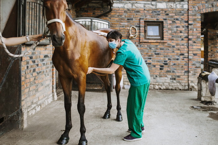
Esophageal obstruction can usually be diagnosed based on clinical signs. Physical examination findings compatible with esophageal obstruction include nasal discharge of feed material and water, bruxism, ptyalism, and palpable enlargement of the esophagus. In some instances, foreign objects lodged in the cervical esophagus may be located via palpation. Subcutaneous emphysema, cervical cellulitis, and fever may be associated with esophageal rupture. The inability to pass a stomach (ruminants) or nasogastric tube (horses) can also confirm the diagnosis.
An endoscopic examination helps localize the site of esophageal obstruction, type of obstructing material, and extent of esophageal ulceration. Because of the risk of aspiration pneumonia, the respiratory tract should be evaluated carefully, including auscultation of the heart and lungs and thoracic radiography. In complicated or chronic cases, a CBC and serum biochemistry profile should be performed. Suggestive CBC abnormalities include leukocytosis, left shift, toxic neutrophils, and hyperfibrinogenemia, whereas biochemical abnormalities include hyponatremia, hypochloremia, and hypokalemia secondary to excessive loss of saliva.
Treatment of Esophageal Obstruction in Large Animals
Sedatives and muscle relaxers to relax the esophagus
Passage of oro- or nasogastric tube to relieve esophageal obstruction
Antimicrobial and anti-inflammatory medications
In horses, many cases of esophageal obstruction may resolve spontaneously if feed and water are withheld. Spontaneous resolution can be aided by IV administration of sedatives (eg, xylazine and detomidine). Oxytocin (0.11–0.22 U/kg, IV, once) is useful to relax esophageal smooth muscle. To ensure that the esophageal obstruction has resolved completely, all horses with suspected obstruction should have a nasogastric tube passed into the stomach or an endoscopic examination.
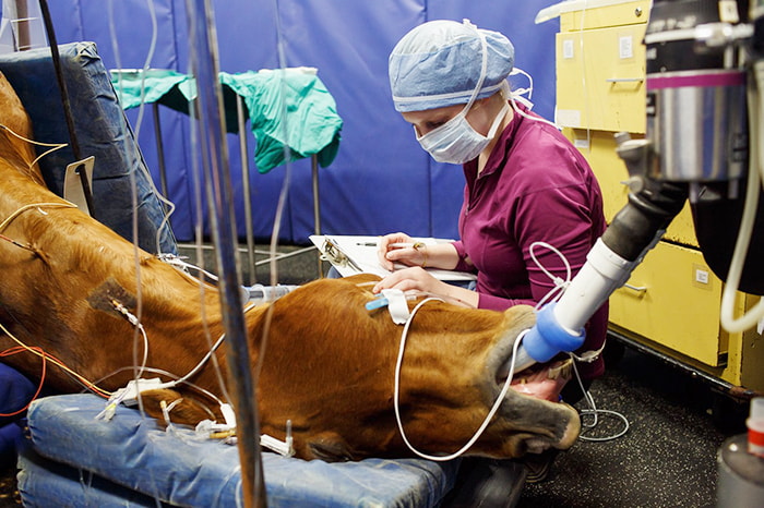
For horses unresponsive to standing esophageal lavage, general anesthesia should be considered, with the horse positioned in lateral recumbency and orotracheally intubated. Again, the head must be positioned lower than the torso to prevent water passing into the lungs. A cuffed endotracheal tube (18–22 mm) is inserted into the esophagus as far as possible or to the level of the esophageal obstruction, and the cuff inflated. A nasogastric tube is inserted through the endotracheal tube, and the esophagus is lavaged. Again, resolution of obstruction should be confirmed with endoscopy or passage of the nasogastric tube into the stomach. An esophagotomy to resolve esophageal obstruction is rarely required.